Early detection is the key to winning the fight against breast cancer. That’s why the American Cancer Society recommends annual breast cancer screenings starting as early as age 40. Different tests are available to help find cancer before you exhibit any outward symptoms. Mammograms are the standard screening method.
There are two types of mammograms. First is the conventional version, which takes a two-dimensional image of each breast to look for any abnormalities. The second more advanced breast imaging technology is called 3D mammography. This uses several x-ray images taken from different angles to create a three-dimensional picture.
3D mammography is FDA-approved, but is relatively new, and isn’t offered at all hospitals and mammogram facilities yet. Salem Radiology Consultants is pleased to offer 3D mammography to our patients in the Salem and the surrounding communities .
How Does3D Mammography Work?
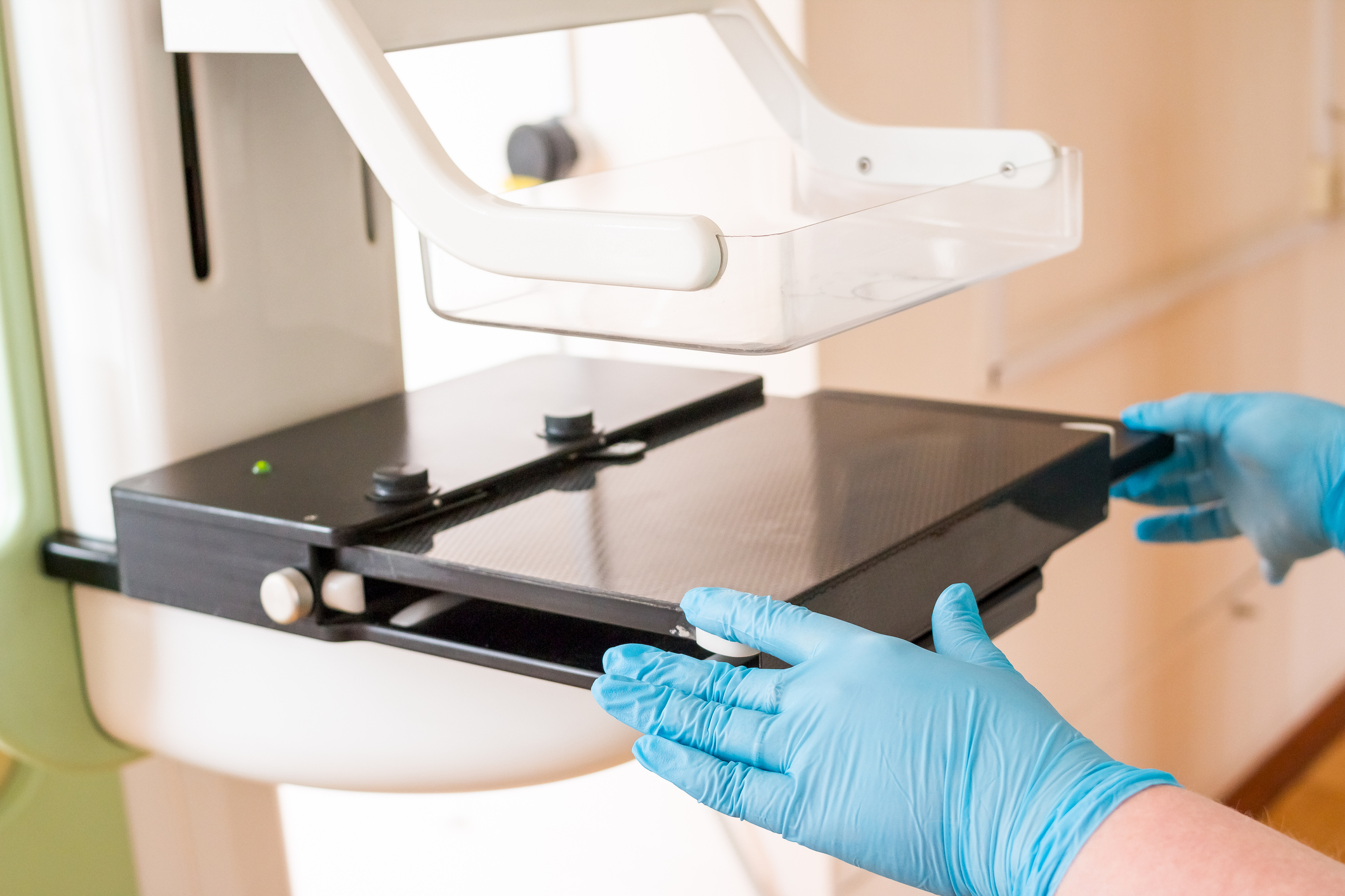
When you visit our clinic for a 3D mammogram, a female technologist helps to position you in front of the machine, which gently compresses your breast to hold it stable during the procedure. Then, an X-ray tube moves in an arc across the breast, capturing 11 images in just seven seconds. The information is sent to a specially designed computer where a physician radiologist carefully examines the highly focused, 3D images of your breast tissue.
Why Choose 3D Mammography?
Consider the benefits of this superior breast screening technology as outlined in Journal of the American Medical Association (JAMA).
Reduced Callbacks
If the doctor detects abnormalities in your mammogram, you may be called back to perform additional imaging. 3D mammography reduces callbacks by 40% eliminating the stress, time, and additional costs associated with follow-up imaging and potential biopsies.
Enhanced Cancer Detection
Screening with 3D mammography leads to a 41% increase in invasive cancer detection compared to conventional mammograms.
Earlier cancer detection
A 2D screening is akin to standing at the edge of a forest and looking for a bird somewhere inside. A 3D screening, on the other hand, is like walking into the forest and looking around 10 steps at a time. The more in-depth nature of 3D imaging allows doctors to detect breast cancer an average of 15 months earlier, giving patients a critical head start in the battle against cancer.
Computerized Second Opinion
The computers used in 3D mammography employ a sophisticated analysis system called computer-aided detection (CAD). After examining the images closely, radiologists consult the CAD system before issuing their final report. This combination of human interpretation and computer analysis increases early breast cancer detection by as much as 19%.
Improved Images in Dense Breast Tissue
Breasts consist of milk glands, milk ducts, and supportive tissue. Breasts are considered “dense” if they contain more supportive tissue than fatty glands or ducts. Dense tissue and cancer both appear as white spots on a standard mammogram, which makes cancer more difficult to detect in dense breasts. However, with 3D mammography, doctors can see beyond areas of density from a different angle, making it easier to detect the presence of cancer and reducing the number of false positives.
More Comfortable Screening
The breast compression required during a conventional mammogram is uncomfortable for many women, causing some women to delay scheduling their exam. Compression also causes some tissue to overlap, giving breast cancer a place to hide. 3D mammography machines only apply enough pressure to keep the breast in a stable position. This makes the procedure much more comfortable without detracting from its effectiveness.
According to one of the senior authors of the study published in JAMA, “These findings reaffirm that 3D mammography is a better mammogram for breast cancer screening. These results are an important step leading toward updated policies so that all women can receive 3D mammography for screening.”
Limitations of 3D Mammography
While this technology provides the safest, and most effective way to screen for breast cancer, it does have a few technological limitations that patients should know about.
Low-Level Radiation Exposure
3D imaging using multiple x-rays to create a three-dimensional image, which exposes patients to low levels of radiation. 2D mammograms use x-rays as well, but because 3D screenings are often combined with standard mammograms, the overall radiation exposure may be slightly higher than using 2D technology alone. However, Salem Radiology’s 3D machines create both two- and three-dimensional images at once, lowering the amount of overall radiation.
Unable to Detect All Cancers
While 3D mammography is better at finding cancerous growths earlier than 2D screenings, small or difficult-to-see tumors may not be detected by the technology. That’s why, no matter what type of imaging you choose, you should continue to schedule breast cancer screenings every year and visit your doctor for a physical breast exam.
Schedule 3D Mammography in Salem, OR
Salem Radiology Consultants is committed to offering the best medical screenings and diagnostic services to our patients. As such, we are proud to be among the early adopters of 3D mammogram technology.
In addition to 3D mammography, we also offer additional imaging techniques, including breast MRIs, breast ultrasounds, and ultrasound-guided breast biopsies. Whether you need preventative screenings or diagnostic services, we can meet your needs at our imaging facility.
If you still have questions, or you’re ready to schedule a 3D mammogram, please contact Salem Radiology Consultants at (503) 399-1262.
